|
What you need—plus what you’ll love MelanieTM software has been your partner in 2D gel and blot image analysis for 35+ years. Whether for detecting subtle protein expression changes or categorizing proteins as present or absent, Melanie has consistently delivered precision and reliability. Melanie 9.3 builds on the features you already love while introducing transformative tools to elevate your analysis. It reinvents the way you approach 2D gel and blot analysis, introducing practical innovations to streamline your workflows, empower collaboration, and ensure traceability and compliance, while further expanding its industry-leading visualization tools. Each new feature has been crafted to solve real research challenges, making your analysis easier, faster and more insightful than ever before. |
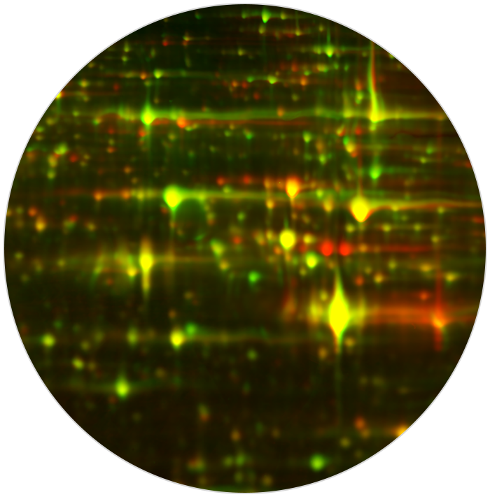
Multichannel color overlay |
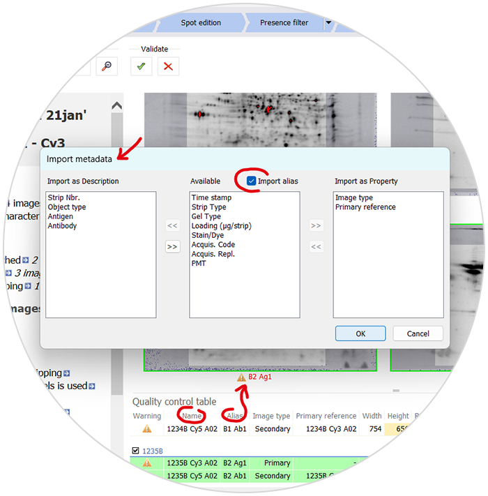
Image aliases and metadata import |
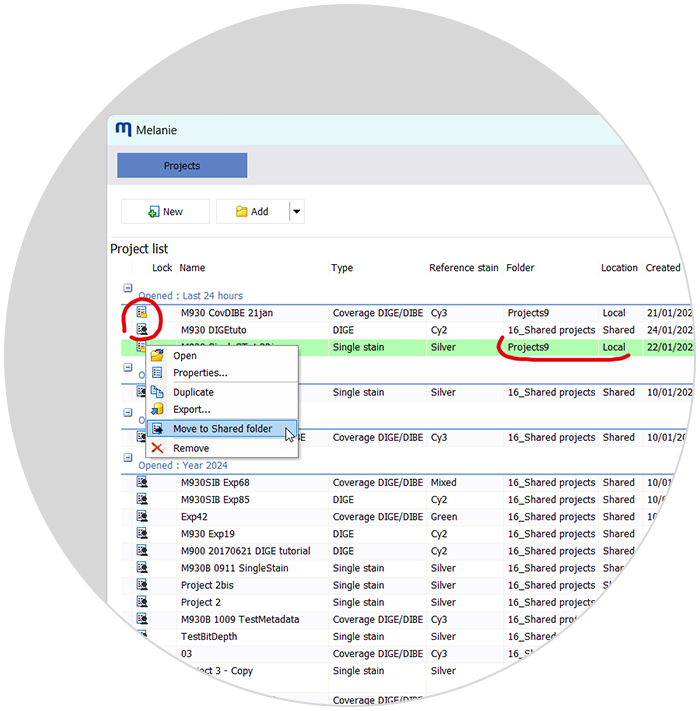
Shared projects and concurrent access management |
| See your protein expression data like never before. The multichannel color overlay lets you blend multiple images with your preferred color scheme to easily visualize similarities and differences between gels and blots. Overlay up to three images in color or any number in grayscale. They don’t need to be from the same gel or blot, as long as they’re aligned. Jazz up your presentations and publications with this powerful display. | Say goodbye to limiting file names. Image aliases allow you to assign clear, descriptive names to images while preserving the original file names. Want to rely on image metadata for analysis? Import sample details, experimental parameters, acquisition dates and more from Excel for seamless integration into your workflow. Spend less time renaming and manually entering information, and more time analyzing. | Collaboration just got easier. Store projects in a shared network folder, ensuring team members always have access to the latest data. Melanie’s smart access management prevents accidental overwrites—if someone else is editing, you’ll get a clear notification. Work securely and efficiently, whether in a small team or a large research group. |
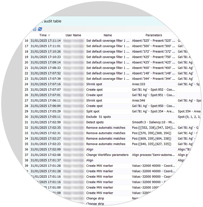
Audit trails and data integrity checks |
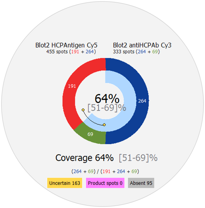
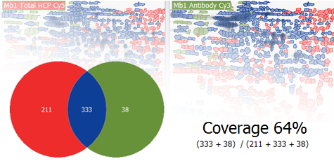
New coverage tools |
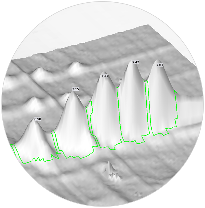
Labels in 3D views and more… |
| Keep your analysis transparent, traceable, and secure. Each action is recorded with timestamps, user IDs, and key parameters for complete auditability, and automatic integrity controls flag external tampering. While full compliance with FDA 21 CFR Part 11 requires additional process and system controls, Melanie provides a solid foundation for data confidence in your operations. | Gain deeper insights into antibody performance. The coverage counter diagram provides a clear, at-a-glance view of coverage results and spot categories. Coverage by volume offers an alternative metric for assessing antibody reactivity when spot count percentages are similar, helping identify the antibody that binds more effectively to high-abundance spots. Make informed reagent choices and ensure more reliable ELISA validation. | Spot labels are now available in 3D view, which further expands the presentation of results, especially for charge variant analysis.
Melanie 9.3 supports even more input image formats, has been validated for Windows 11 and includes automatic update notifications. Plus, a host of usability improvements and bug fixes make this version more robust than ever. |
| Learn more about Melanie 9.3 |
|
Supported workflows Melanie lets you perform all your gel-based protein expression profiling with one simple, comprehensive solution so that you do not need to install, maintain or master several software packages. |
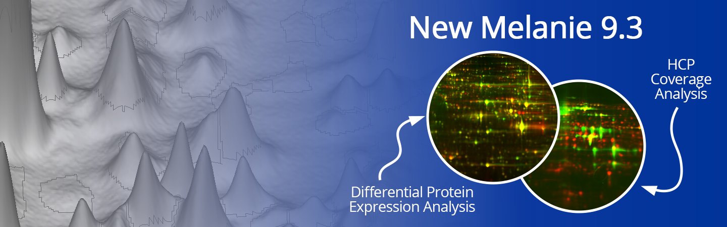
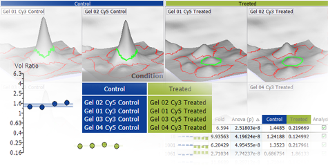
Differential Protein Expression Analysis |
HCP Antibody Coverage Analysis |
|
|
|
|
|
| For scientists investigating biological processes by characterizing samples in terms of relative protein abundance and post-translational modifications. | For scientists involved in the development or use of immunoassays aimed at monitoring Host Cell Protein (HCP) impurities in biopharmaceutical products. | |
| Designed to detect statistically significant differences in protein expression between experimental groups, with high objectivity, sensitivity and confidence. | Designed to analyze 2D-PAGE based coverage, i.e. the percentage of immunodetection that an antibody reagent offers for the total population of HCPs. | |
| Supports images from a wide range of 2D-PAGE based methods: • Conventional 2-DE • 2D Differential in Gel Electrophoresis (2D-DIGE) • Other multiplexed technologies without internal standard |
Supports images from a wide range of 2D-PAGE based methods: • Conventional 2-DE followed by 2D Western blotting • 2D Differential In Blot Electrophoresis (2D-DIBE) • Immunoaffinity chromatography followed by 2D-DIGE |
|
| Learn more | Learn more |
Not sure what solution is most appropriate for your needs?Then learn more here. Or book a free discovery session. An application specialist can clarify the differences between Melanie’s version and licensing options, show you the software at work, explain how to obtain a trial license to evaluate Melanie with your own gel or blot images, and provide recommendations and tips to quickly get started with your analysis.
|
|
| Melanie Classic and DIGE | Melanie Coverage |
| for differential expression analysis | for HCP coverage analysis |
Ask your question
|
|
Guided step-by-step workflow |
Unequaled flexibility in image visualization |
Supports all common detection methods and image formats |
Free Viewer functionality |
|||
| Melanie offers significant time savings with the intuitive step-by-step workflow. Tools dedicated to each analysis stage let you feel confident that you accomplish the necessary steps and checks for the highest quality results. | Melanie gives you full liberty over how you want to group and layout your images for viewing, and lets you choose your preferred combination of 2D and/or 3D views. No other software allows you to adjust the specific views with such ease and flexibility. | Melanie supports any type of staining, including colorimetric, fluorescence and functional group specific stains, as well as DIGE images or western blots. Similarly, all common image formats can be analyzed, including TIFF, GEL, MEL, IMG, GSC, 1SC and SCN files. | Melanie lets you open your gel images and check their quality even without a license. Any collaborator will be able to view the results of an analysis carried out with the licensed software, so you can share your work and scientific discoveries. Installing the license will unlock all functionality. |