Image alignment demystified: Part 3 – Recommendations for successful alignment
Welcome back to our “Image Alignment Demystified” article series. In part 1, we took a deep dive into why alignment is pivotal in 2D gel electrophoresis analysis and explored its fundamental principles. Part 2 focused on the criteria and tools to help you assess alignment quality. In this chapter, we continue our journey by offering a practical roadmap with 10 recommendations and guidelines to ensure you achieve the best alignment results, thereby maximizing the reliability and precision of spot correspondences.
1. Clear the deck: Purge doubtful matches
The journey to accurate alignment begins with clearing the deck. Doubtful matches, which we define as those that seem incorrect, uncertain, or lack clear alignment evidence, should be removed. This step allows you to concentrate on establishing stronger, more trustworthy spot correspondences.
2. The gold standard: Only match clearly corresponding spots
The gold standard in alignment is matching only those spots that you’re certain represent the same protein. Be careful not to match different protein variants or spots that do not represent identical protein isoforms. The crux of proficient matching lies in identifying distinctive, unambiguous features that signal spot correspondence.
3. Precision alignment: Target mid-sized and small spots
When manually adding matches, your best bet is mid-sized or small spots with a defined peak. Large or saturated spots pose challenges in identifying the precise peak position, potentially introducing inaccuracies into the alignment process.
4. Moderation over extremes: Rethinking overzealous matching
It might be tempting to match every spot manually, but this practice isn’t necessary or even desirable. The raison d’être of both automatic and user-defined matches is to achieve a flawless overlap, where the aligned image is perfectly superimposed on the reference image. Gels with minimal positional variation may require few matches to achieve that. So exercise restraint and avoid overcomplicating the process.
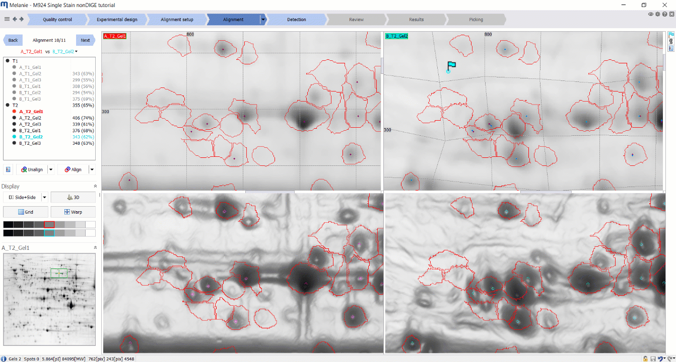
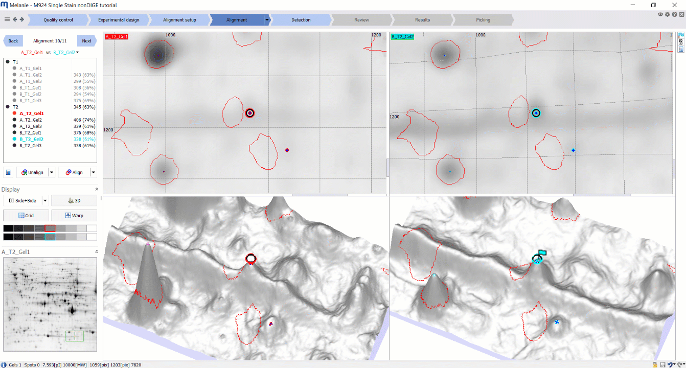
5. Positional parity: Harmonize positions for precision
Paying attention to the exact positions of the matches is another cornerstone of successful alignment. While the matches don’t have to be at the very top of each spot, the positions should mirror each other in both images. This practice ensures alignment accuracy and reduces positional discrepancies. Moreover, matches need not be restricted to ‘detected’ signals marked with a spot outline; any pixel within an image can be matched with a corresponding pixel on the reference image.
6. Steer clear of ambiguity: Navigate ambiguous waters with care
Certain situations can muddy the waters – a single spot peak in one image corresponding to multiple peaks in another is a classic example. When in doubt, it’s always better to err on the side of caution. Avoid creating a match or remove any automatic matches that appear ambiguous.
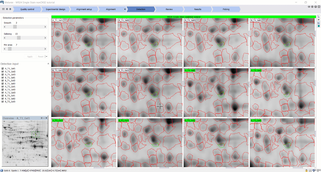
7. Special strategy: Tackle dense or highly variable regions
Highly variable regions or areas with dense spot patterns, including those with artifacts or overlapping charge variants, demand a special strategy. Matching spots around these areas usually proves more effective than matching spots within them. This counter-intuitive approach often leads to superior alignment outcomes. Of course, when confidence in corresponding protein isoforms is high, exceptions can be made, with the 3D view offering crucial assistance.
8. Fresh start: The clean slate approach
Confronted with challenging alignments featuring numerous incorrect automatic matches? Don’t hesitate to start afresh. Unaligning and then manually creating a few matches distributed across the gel can work wonders. Begin by matching two clearly corresponding spots, and expand your match territory from there. Keep in mind that if your brain cannot detect similarities in the spot pattern, the algorithm will likely fail as well. When it doesn’t, treat automatic results with an extra degree of scrutiny.
9. Climb the ladder: Keep the alignment hierarchy in mind
In scenarios employing multi-level alignment strategies, peculiar shifts in certain spot shapes might appear despite the current alignment seeming accurate. These oddities can hint at misalignments higher up the ladder. It’s paramount to revisit and rectify these higher-level alignments to maintain spot-matching accuracy throughout the process.
10. Pre-emptive action: The alignment-detection interplay
Before embarking on the intricacies of spot editing or annotation, take a moment to evaluate the spot pattern in the Detection or Review steps. In particular, look for signs such as shifted spot contours relative to certain peaks, or spot shapes that are very different from those in the reference image. Such indicators can point to misalignments and signal that you may need to loop back to the Alignment step. Taking a proactive stance on alignment discrepancies will not only save you tedious corrections down the line, but will also safeguard your analysis from misleading conclusions.
Closing thoughts
The alignment of images stands as the cornerstone of 2D gel electrophoresis analysis. By diligently adhering to the recommendations outlined above, you can streamline the alignment process and achieve accurate, reliable, and precise spot matching, leading to successful image analysis. As we wrap up this instalment, we look forward to discussing further insights in the final part of our “Image Alignment Demystified” series, where we will discuss some final considerations to keep in mind.